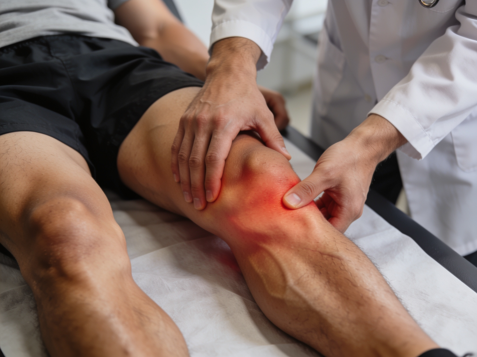
Knee pain that is deep and aching, worsens with weight-bearing activity, and is accompanied by swelling that keeps recurring may indicate damage to the articular cartilage lining the joint. Cartilage injuries are common in sport, are frequently found alongside ACL and meniscal tears, and require early assessment to prevent progressive joint damage
What is Knee Cartilage Injuries?
Articular cartilage is the smooth white tissue covering the ends of the bones within the knee joint. It allows frictionless movement and absorbs impact. Unlike most tissues in the body, articular cartilage has very limited natural healing capacity because it has no blood supply. This means that unmanaged cartilage defects tend to progressively enlarge and ultimately accelerate the development of knee arthritis.
Types of Knee Cartilage Injuries
Type I
Grade I (Superficial): Softening and surface irregularity of the cartilage without a full-thickness defect. The cartilage surface shows blistering or fibrillation but remains continuous. Often an incidental finding on MRI or arthroscopy and may not require specific surgical treatment.
Type II
Grade II (Partial Thickness): A defect extending less than half the depth of the cartilage. The bone is not exposed. Partial thickness defects can be painful and progressive, particularly in younger active patients.
Type III
Grade III (Deep Partial to Full Thickness): A defect extending to more than half the cartilage depth, reaching toward the subchondral bone but not fully breaching it. Grade III lesions cause significant pain with loading and are associated with progressive joint degeneration if left untreated.
Type IV
Grade IV (Full Thickness): Complete loss of cartilage exposing the subchondral bone. The bone surface is visible at the base of the defect. Grade IV lesions cause bone-on-bone contact during weight-bearing and are associated with significant pain and accelerated development of knee arthritis.
Type V
Osteochondral Defect: An injury involving both the overlying articular cartilage and the underlying subchondral bone, often resulting from trauma or osteochondritis dissecans. A fragment of bone and cartilage may be partially or completely detached, creating a loose body within the knee joint.
Causes
Acute traumatic impact during sport, such as a direct blow to the knee from a collision, a landing from height, or a twisting injury with an axial compressive component, can shear a fragment of cartilage from the underlying bone in a single event.
Chronic repetitive loading from sport and physical activity gradually wears the cartilage surface over time, particularly in areas of high contact stress such as the medial femoral condyle. Runners, footballers, and kabaddi players are among the most commonly affected groups in Bangalore.
Co-existing joint pathology including ACL instability, meniscal tears, and lower limb malalignment alter the pattern of contact stress within the knee and accelerate cartilage wear in specific locations. It is common to find cartilage damage alongside ACL and meniscal injuries at the time of arthroscopy.
Osteochondritis dissecans, a condition in which a segment of subchondral bone loses its blood supply, is a specific cause of osteochondral defects, most commonly affecting the lateral aspect of the medial femoral condyle in adolescents and young adults.
Inflammatory conditions including rheumatoid arthritis directly attack and erode articular cartilage through enzymatic destruction of the cartilage matrix, producing diffuse rather than focal cartilage loss.
- Deep, aching knee pain that worsens with weight-bearing activities such as walking, running, climbing stairs, and squatting
- Swelling of the knee that develops after activity and persists into the following day, often described as the knee feeling puffy or heavy after exercise
- Stiffness particularly after sitting for prolonged periods or after rest, improving with gentle initial movement then worsening again with continued loading
- A catching, clicking, or locking sensation if a loose osteochondral fragment is present and moving freely within the joint
- Giving way of the knee, particularly in patients with associated ligamentous instability or when an osteochondral fragment causes sudden pain during a movement
- Pain localised to a specific compartment of the knee, either medial, lateral, or anterior, corresponding to the area of cartilage damage
- In advanced cases, persistent pain at rest and at night as the cartilage loss progresses toward full-thickness bone-on-bone contact

Diagnosis
Clinical examination identifies the area of maximal tenderness, assesses for joint effusion, and tests for associated ligamentous and meniscal pathology that frequently accompanies cartilage injury. However, articular cartilage damage does not produce specific clinical signs that reliably distinguish it from other intra-articular pathology, making imaging essential for an accurate diagnosis.
MRI is the gold standard investigation for knee cartilage assessment, providing detailed information on cartilage thickness, signal intensity, the presence of subchondral bone oedema or cysts, and any associated osteochondral fragments. Dedicated cartilage-sensitive MRI sequences provide quantitative assessment of cartilage quality and help grade the severity of the defect. Plain X-rays may show joint space narrowing in advanced cartilage loss but are frequently normal in early and moderate disease, making MRI the preferred investigation when cartilage injury is suspected. Diagnostic arthroscopy remains the most accurate method of directly visualising and grading cartilage defects, and allows treatment to be performed in the same operative session when appropriate.
Treatment
Arthroscopic Microfracture
For smaller focal cartilage defects with intact surrounding cartilage, Dr. Kushalappa performs microfracture, an arthroscopic technique where small perforations are made in the underlying bone to stimulate a healing fibrocartilage response from bone marrow stem cells. This is a first-line procedure for focal defects under 2 to 3 cm in size.
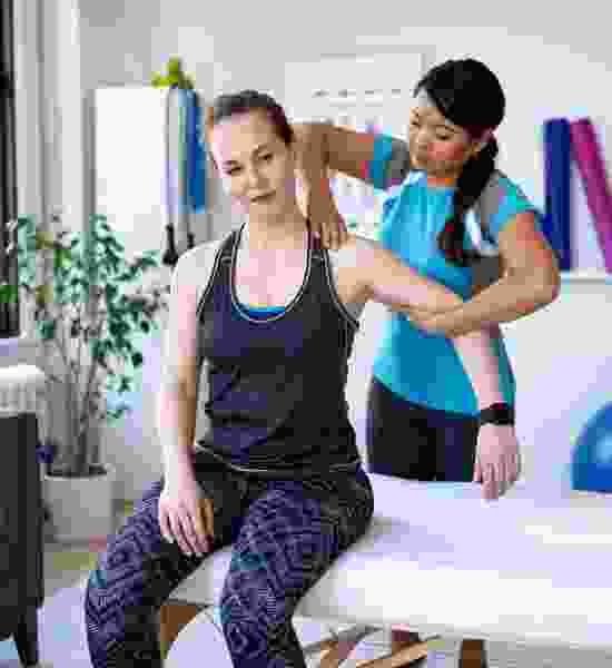
Why Choose Dr. Kushalappa Subbiah in Bangalore?
Dr. Kushalappa Subbiah completed a Fellowship in Shoulder Surgery at the Sydney Shoulder Research Institute, where he trained in advanced arthroscopic shoulder procedures including labral repair and reconstruction. He holds the International Olympic Committee (IOC) Diploma in Sports Medicine, and has direct clinical experience managing shoulder injuries in Indian athletes across cricket, swimming, tennis, and contact sports. He is appointed as a Consultant Shoulder Surgeon at NH Hospital, Bangalore.
Frequently Asked Questions
Articular cartilage has very limited capacity for natural regeneration. Microfracture produces a fibrocartilage repair tissue that is biomechanically inferior to native hyaline cartilage but adequate for most functional demands. More advanced techniques such as cartilage transplantation are available for larger defects.