A sudden sharp pain below the kneecap during a jump or explosive push-off movement, combined with an immediate inability to straighten the knee and a palpable defect in the front of the knee, is the presentation of a patellar tendon rupture. It requires prompt surgical repair to restore full knee extensor function.
What is Patellar Tendon Rupture?
The patellar tendon connects the patella (kneecap) to the tibia (shinbone) and transmits the power of the quadriceps muscle to produce knee extension. It is subject to very high forces during jumping and sprinting. Complete ruptures can occur in athletes during explosive movements, or in individuals with pre-existing tendinopathy where the tendon has been progressively weakened.
Types of Patellar Tendon Rupture
Type I
Acute Complete Rupture: The entire patellar tendon tears, most commonly at its proximal attachment to the inferior pole of the patella. The patient cannot actively extend the knee and a palpable gap is present below the kneecap. Requires urgent surgical repair.
Type II
Acute Partial Rupture: Only a portion of the patellar tendon fibres are disrupted, preserving some knee extension function. The patient may retain the ability to extend the knee against gravity but with significant weakness and pain. MRI is required to characterise the extent of the tear and guide treatment.
Type III
Rupture Through a Degenerate Tendon: Complete rupture occurring spontaneously or with minimal force in a tendon that has been chronically weakened by patellar tendinopathy. A subset of patients with longstanding jumper's knee ultimately experience spontaneous tendon failure. The underlying degeneration is identified on MRI and must be accounted for in surgical planning.
Type IV
Avulsion Fracture at the Tibial Tuberosity: In skeletally immature patients, the equivalent of a patellar tendon rupture may present as an avulsion fracture of the tibial tuberosity, where the tendon pulls off a bony fragment rather than tearing through the tendon substance itself.
Causes
An unexpected eccentric load through the quadriceps mechanism is the most common mechanism. This typically occurs when an athlete misjudges a landing from a jump and the knee is driven into flexion while the quadriceps contracts violently to resist it.
Pre-existing patellar tendinopathy weakens the structural integrity of the tendon and significantly increases the risk of rupture, sometimes occurring with a relatively minor load that a healthy tendon would easily withstand.
Systemic conditions including chronic renal failure, diabetes mellitus, hyperparathyroidism, and prolonged corticosteroid use predispose tendons to spontaneous rupture by impairing collagen synthesis and repair.
Local corticosteroid injections directly into the patellar tendon are strongly associated with subsequent tendon rupture and should be avoided in patellar tendinopathy management.
- A sudden sharp, tearing pain at the front of the knee, typically felt just below the kneecap during a jump, sprint, or sudden deceleration
- An audible pop or snap at the moment of rupture, heard and felt by the patient
- Immediate inability to straighten the knee or bear weight on the leg following the injury
- A visible or palpable gap immediately below the kneecap, where the patellar tendon has torn away from the inferior pole of the patella
- The kneecap riding abnormally high, called patella alta, as the proximal retraction of the torn tendon allows the patella to migrate superiorly
- Rapidly developing swelling and bruising around the front of the knee

Diagnosis
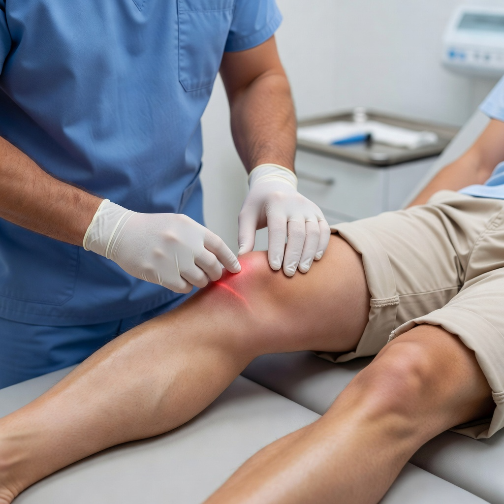
Clinical examination is often sufficient to confirm a complete patellar tendon rupture. The combination of inability to extend the knee against gravity, a palpable gap below the kneecap, and a superiorly positioned patella on palpation is highly diagnostic. In partial tears where some extension is retained, the clinical picture is less clear and imaging becomes essential.
Plain X-rays demonstrate a high-riding patella, which is a reliable radiographic sign of complete rupture. A small bony fragment avulsed from the inferior patellar pole may also be visible. Ultrasound provides rapid dynamic assessment of the tendon and can confirm the gap and degree of retraction. MRI is used to confirm partial tears, assess the degree of retraction in complete ruptures, and identify any co-existing pathology such as patellar tendinopathy or bone bruising that will influence the surgical approach.
Treatment
Surgical Repair
Complete patellar tendon ruptures require prompt surgical repair. Dr. Kushalappa reattaches the ruptured tendon to the tibial tuberosity using non-absorbable sutures and bone anchors through an anterior knee approach. Early repair within 1 to 3 weeks of injury is strongly preferred. Recovery involves a period of immobilisation in extension for 4 to 6 weeks, followed by progressive physiotherapy. Return to full sport is expected at 9 to 12 months.
Why Choose Dr. Kushalappa Subbiah in Bangalore?
Dr. Kushalappa Subbiah completed a Fellowship in Shoulder Surgery at the Sydney Shoulder Research Institute, where he trained in advanced arthroscopic shoulder procedures including labral repair and reconstruction. He holds the International Olympic Committee (IOC) Diploma in Sports Medicine, and has direct clinical experience managing shoulder injuries in Indian athletes across cricket, swimming, tennis, and contact sports. He is appointed as a Consultant Shoulder Surgeon at NH Hospital, Bangalore.
Frequently Asked Questions
Surgical repair should ideally be performed within 2 to 3 weeks of the injury. Early repair allows direct suturing of the tendon ends without the need for reconstruction grafts. Delays result in tendon retraction and scarring, making repair technically more difficult and recovery longer.