A sudden, severe pain above the kneecap during a fall, a jump, or a sudden forced knee bend, combined with an inability to straighten the leg and a visible or palpable defect above the patella, is the presentation of a quadriceps tendon rupture. It tends to occur in patients over 40 and demands prompt surgical repair.
What is Quadriceps Tendon Rupture?
The patellar tendon connects the patella (kneecap) to the tibia (shinbone) and transmits the power of the quadriceps muscle to produce knee extension. It is subject to very high forces during jumping and sprinting. Complete ruptures can occur in athletes during explosive movements, or in individuals with pre-existing tendinopathy where the tendon has been progressively weakened.
Types of Quadriceps Tendon Rupture?
Type I
Complete Rupture at the Superior Patellar Pole: The most common pattern. The quadriceps tendon tears entirely from its attachment at the top of the patella. The quadriceps muscle belly retracts proximally, producing a visible suprapatellar fullness and a palpable gap above the patella. This pattern requires surgical repair.
Type II
Partial Rupture: Only a portion of the quadriceps tendon fibres are torn, most commonly the rectus femoris layer, with the deeper vastus medialis and lateralis fibres remaining intact. Knee extension against gravity is preserved but with significant weakness and pain. MRI is required to quantify the extent of the tear and guide the treatment decision.
Type III
Bilateral Simultaneous Rupture: An uncommon but significant presentation in which both quadriceps tendons rupture simultaneously, typically in the context of systemic predisposing conditions such as chronic renal failure or hyperparathyroidism. Should prompt thorough medical investigation for underlying metabolic bone disease.
Causes
An unexpected forceful eccentric contraction of the quadriceps, most commonly occurring when stumbling or when a patient with reduced balance and proprioception misjudges a step, causes the tendon to fail at its insertion on the patella.
Pre-existing tendon degeneration, which is extremely common in patients over 40 with systemic conditions, significantly predisposes the quadriceps tendon to failure at loads far below those that a healthy tendon would easily tolerate.
Chronic renal failure on dialysis is one of the most strongly associated systemic risk factors. Renal patients have elevated levels of secondary hyperparathyroidism, amyloid deposition in tendons, and altered collagen metabolism that collectively weaken tendon integrity.
Corticosteroid use, whether systemic for inflammatory conditions or inappropriately injected into the tendon, impairs collagen synthesis and significantly increases rupture risk.
Diabetes mellitus, obesity, and other systemic metabolic conditions are recognised predisposing factors through their effects on tissue vascularity and collagen quality.
- A sudden severe pain above the kneecap at the moment of rupture, often described as a tearing or snapping sensation
- Immediate inability to straighten the knee against gravity, producing a characteristic flexed knee posture and an inability to walk normally
- The patella sitting lower than normal, called patella baja, as the proximal quadriceps pull has been lost and the patella descends under the influence of the intact patellar tendon
- A visible or palpable gap or indentation directly above the patella, where the quadriceps tendon has torn away from the superior patellar pole
- Rapid swelling and bruising of the anterior knee and lower thigh
- In bilateral ruptures: inability to rise from a chair or climb stairs without upper limb support, with both legs giving way simultaneously when weight is transferred onto the legs

Diagnosis
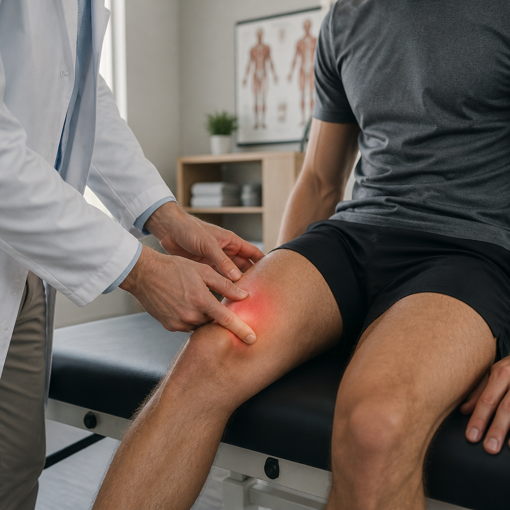
Clinical examination confirms the diagnosis in complete ruptures. Inability to perform a straight leg raise or extend the knee against gravity, a palpable gap above the patella, and a low-riding kneecap on palpation are the defining clinical features. In the setting of acute swelling, the gap may be partially obscured and careful palpation is required.
Plain X-rays demonstrate a low-riding patella and may show a small bony avulsion fragment at the superior patellar pole. Ultrasound provides immediate bedside assessment and can confirm the gap and degree of retraction. MRI is the gold standard investigation for confirming partial tears, measuring the degree of retraction in complete ruptures, characterising the degree of pre-existing tendon degeneration, and identifying any associated pathology at the patellar bone-tendon interface that will affect surgical planning.
Treatment
Surgical Repair
Complete quadriceps tendon ruptures require surgical repair. Dr. Kushalappa reattaches the torn tendon to the superior patella using sutures and anchors through an open anterior approach. As with patellar tendon repair, early surgery within 2 to 3 weeks provides the best outcomes. Recovery involves immobilisation for 4 to 6 weeks, physiotherapy over the following months, and return to full activity at 9 to 12 months.
Why Choose Dr. Kushalappa Subbiah in Bangalore?
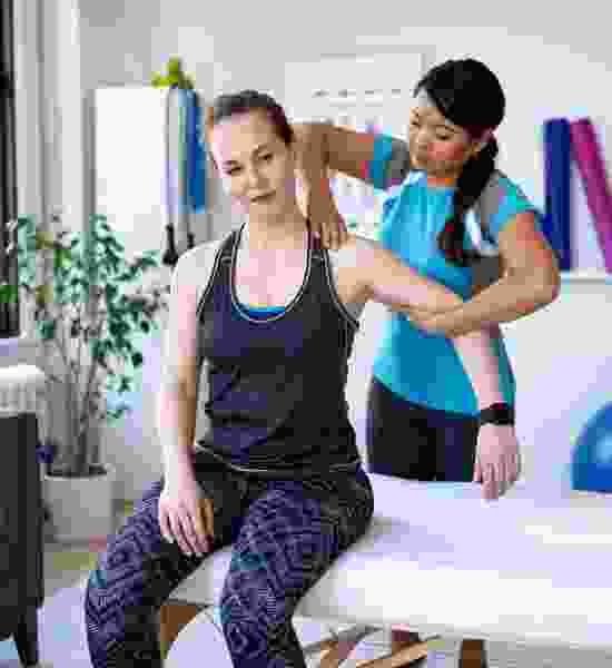
Dr. Kushalappa Subbiah completed a Fellowship in Shoulder Surgery at the Sydney Shoulder Research Institute, where he trained in advanced arthroscopic shoulder procedures including labral repair and reconstruction. He holds the International Olympic Committee (IOC) Diploma in Sports Medicine, and has direct clinical experience managing shoulder injuries in Indian athletes across cricket, swimming, tennis, and contact sports. He is appointed as a Consultant Shoulder Surgeon at NH Hospital, Bangalore.
Frequently Asked Questions
Yes, particularly in the acute setting where significant swelling can mask the palpable gap above the patella. The key clinical finding is inability to actively extend the knee against gravity, which should prompt an urgent orthopaedic referral and MRI confirmation.