6935 Todds Lane San Antonio, TX 98336
Our Working Time: 09:00 am To 06:00 pm

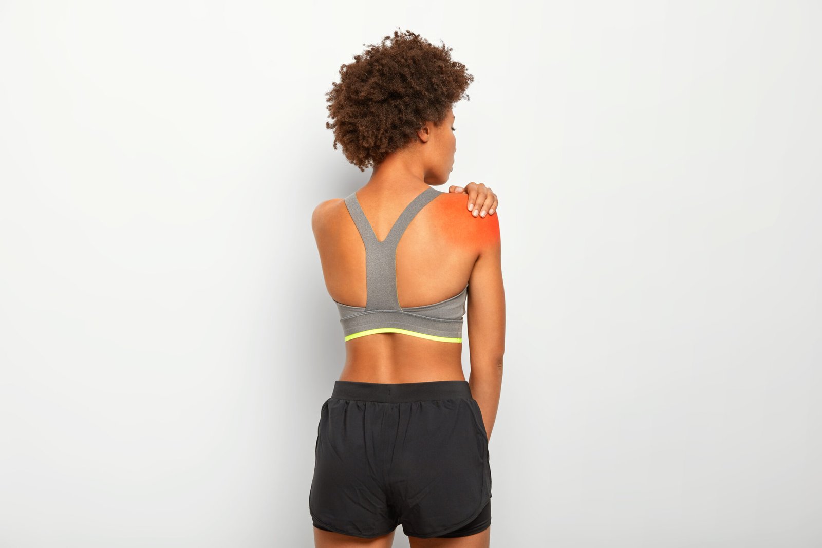
A fall onto the shoulder or an outstretched arm, a tackle in football or rugby, or a road traffic accident, any of these can fracture the clavicle, the collarbone that connects the shoulder blade to the breastbone. It is one of the most common fractures in children and young adults and, when managed well, most patients make a full recovery.
What is Clavicle Fracture?
The clavicle is the slender S-shaped bone that runs horizontally between the sternum and the acromion of the shoulder blade, forming the front strut of the shoulder girdle. Clavicle fractures account for approximately 10 percent of all fractures and are among the most common injuries in contact sport and cycling. Most fractures occur in the middle third of the bone.
Types of Clavicle Fracture
Type I
Mid-Shaft Fracture (Group I, approximately 80 percent of all clavicle fractures): The fracture occurs in the middle third of the clavicle, the narrowest and most exposed segment of the bone. These fractures range from undisplaced hairline fractures to severely shortened and displaced injuries with multiple fragments.
Type II
Lateral (Distal) Third Fracture (Group II, approximately 15 percent): Fractures near the acromioclavicular joint. Type II lateral fractures, where the coracoclavicular ligaments are disrupted, have a higher non-union rate and are more likely to require surgical intervention.
Type III
Medial (Proximal) Third Fracture (Group III, approximately 5 percent): Fractures near the sternoclavicular joint. Less common but potentially more dangerous due to proximity to the great vessels and trachea. CT scan is important to assess displacement relative to these structures.
Causes
Direct falls onto the shoulder or outstretched arm are the most common mechanism. Contact sport collisions, cycling accidents, and road traffic injuries are other frequent causes.
- Immediate pain along the collarbone following injury
- A visible bump or deformity at the fracture site
- Bruising and swelling over the clavicle
- Reluctance to move the arm, holding it close to the body
- A grinding sensation with any arm movement

Diagnosis
Plain X-rays are diagnostic and guide treatment decisions. CT scan is used for complex fractures near the joints at either end of the clavicle.
Treatment
Non-Surgical Management
The majority of mid-shaft clavicle fractures, particularly undisplaced or minimally displaced ones, are treated with a sling for 4 to 6 weeks followed by physiotherapy. Bone healing is typically complete within 6 to 12 weeks in adults.
Surgical Fixation
Significantly shortened or displaced fractures, particularly those with more than 2 cm of shortening or multiple fragments, are associated with poorer outcomes from conservative management. In these cases, Dr. Kushalappa performs open reduction and fixation with a plate and screws, allowing earlier return of shoulder function and reducing the risk of non-union. Athletes and patients who require early return to activity are also offered surgical fixation.
Why Choose Dr. Kushalappa Subbiah in Bangalore?
Dr. Kushalappa Subbiah completed a Fellowship in Shoulder Surgery at the Sydney Shoulder Research Institute, where he trained in advanced arthroscopic shoulder procedures including labral repair and reconstruction. He holds the International Olympic Committee (IOC) Diploma in Sports Medicine, and has direct clinical experience managing shoulder injuries in Indian athletes across cricket, swimming, tennis, and contact sports. He is appointed as a Consultant Shoulder Surgeon at NH Hospital, Bangalore.
Frequently Asked Questions
Undisplaced fractures typically heal within 6 to 8 weeks in adults, and faster in children. Surgically treated displaced fractures may allow earlier functional recovery and usually return to sport within 3 to 4 months.
Some residual bony prominence at the fracture site is common, even after full healing, particularly with displaced fractures treated non-surgically. Surgical fixation reduces but does not always eliminate this deformity.