6935 Todds Lane San Antonio, TX 98336
Our Working Time: 09:00 am To 06:00 pm

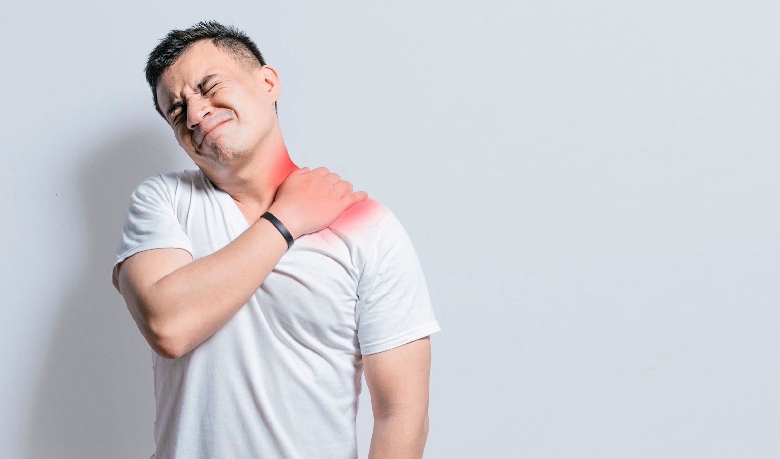
Pain and a visible prominence at the front of the chest near the base of the neck, or a sense that the collarbone has shifted from its normal position, are the key features of a sternoclavicular joint injury. Though less commonly discussed than other shoulder problems, SC joint instability can significantly affect shoulder function and requires specialist assessment to manage correctly.
Dr. Kushalappa Subbiah provides specialist evaluation and management of SC joint conditions at his Bangalore clinics, including both acute injuries and chronic instability presentations.
What is SC Joint?
The sternoclavicular (SC) joint is the articulation between the inner end of the clavicle (collarbone) and the sternum (breastbone) at the centre of the chest. It is the only bony joint connecting the entire upper limb to the axial skeleton. Despite being a small joint, it plays a vital role in transmitting force from the arm to the trunk and allowing the shoulder to move through its full range.
The SC joint is stabilised by strong ligaments on its front and back surfaces, as well as a disc of fibrocartilage within the joint. When these structures are disrupted through trauma or degenerate over time, the joint becomes painful and unstable. SC joint injuries are classified by the direction of displacement of the clavicle: anterior (forward) or the more serious and potentially life-threatening posterior (backward) dislocation.
Types of SC Joint Conditions
Type I
Grade I (Sprain): The SC joint ligaments are stretched but intact. The joint remains stable. Tenderness and mild swelling are present at the joint but no displacement of the clavicle occurs.
Type II
Grade II (Subluxation): Partial tearing of the ligaments causes the clavicle to shift partially out of position but it remains partially reduced. A visible step deformity may be present.
Type III
Grade III Anterior Dislocation: Complete disruption of the SC joint ligaments with the medial clavicle displacing forward in front of the sternum. A visible and palpable prominence is noted at the front of the chest. This is the most common direction of dislocation.
Type IV
Grade III Posterior Dislocation: Complete dislocation with the medial clavicle displaced behind the sternum toward the great vessels and airways. This is a medical emergency requiring urgent assessment, as the clavicle can compress the trachea, oesophagus, or major blood vessels.
Type V
SC Joint Arthritis: Degenerative or inflammatory arthritis of the SC joint produces pain, swelling, and restricted shoulder movement. It may occur as primary osteoarthritis, in association with rheumatoid arthritis, or following previous SC joint injury.
Causes
High-energy trauma is the most common cause of SC joint dislocation. A direct blow to the shoulder or a fall onto the outstretched arm transmits force to the SC joint. Contact sports including rugby, wrestling, and kabaddi are common contexts for anterior SC dislocation. Motor vehicle accidents are a frequent cause of posterior dislocation due to the forces involved. In older patients, degenerative arthritis of the SC joint can develop gradually without a specific traumatic event.
- Pain at the front of the chest at the base of the neck, particularly with shoulder movement, lifting, or lying on the affected side
- A visible or palpable prominence at the SC joint, representing the displaced clavicle end in anterior dislocation
- Swelling and bruising around the joint following an acute injury
- A clicking, grinding, or crepitus sensation with shoulder movement, more common in SC joint arthritis
- In posterior dislocation: difficulty swallowing, shortness of breath, or a choking sensation indicating pressure on adjacent structures — this requires immediate emergency assessment
- Shoulder pain and restricted movement that reduces with rest and worsens with overhead or cross-body activity

Diagnosis
Clinical examination of the SC joint is essential and includes careful assessment of the position of the medial clavicle, local tenderness, and any signs of posterior compression of mediastinal structures. Standard shoulder X-rays are often insufficient to assess the SC joint adequately. CT scan is the investigation of choice for characterising the degree of displacement, identifying associated fractures, and distinguishing anterior from posterior dislocation. MRI is used in chronic presentations where ligament integrity and the intra-articular disc need to be assessed.
Treatment
Non-Surgical Management
Grade I and II sprains are managed conservatively with a sling for comfort, ice, analgesia, and early physiotherapy once the acute pain has settled. Anterior SC dislocations that have been reduced and remain stable with strapping can also be managed non-surgically, though recurrence of instability is not uncommon. Posterior SC dislocations require urgent assessment and often closed reduction under general anaesthesia in a controlled theatre environment due to the risk of vascular and airway injury during the reduction manoeuvre.
SC Joint Reconstruction (Surgical)
For chronic anterior SC joint instability that is symptomatic and has not responded to conservative management, surgical stabilisation is performed. Dr. Kushalappa uses ligament reconstruction techniques using tendon graft to restore stability to the SC joint, protecting the medial clavicle from recurrent displacement. SC joint arthritis that fails conservative management is addressed through resection of the medial clavicle (medial claviculectomy), which reliably resolves pain while preserving shoulder function.
Why Choose Dr. Kushalappa Subbiah in Bangalore?
Dr. Kushalappa Subbiah completed a Fellowship in Shoulder Surgery at the Sydney Shoulder Research Institute, where he trained in advanced arthroscopic shoulder procedures including labral repair and reconstruction. He holds the International Olympic Committee (IOC) Diploma in Sports Medicine, and has direct clinical experience managing shoulder injuries in Indian athletes across cricket, swimming, tennis, and contact sports. He is appointed as a Consultant Shoulder Surgeon at NH Hospital, Bangalore.
Frequently Asked Questions
Anterior SC dislocations, while painful and visually prominent, are generally not life-threatening and can often be managed without surgery. Posterior SC dislocations are a medical emergency because the displaced clavicle can compress the trachea, oesophagus, or major blood vessels behind the sternum. Any patient with a suspected posterior dislocation must be assessed urgently in hospital.
Not all. Acute anterior dislocations may be reduced under sedation or general anaesthesia with gentle pressure. However, chronic dislocations that have settled in a displaced position may not require reduction if they are not causing functional problems. Posterior dislocations nearly always require formal reduction in a theatre setting due to the surrounding vascular and airway structures.
Yes. The initial management of SC joint arthritis includes anti-inflammatory medications, activity modification, physiotherapy, and corticosteroid injections into the joint. Surgery is only considered when symptoms persist despite adequate conservative treatment.