6935 Todds Lane San Antonio, TX 98336
Our Working Time: 09:00 am To 06:00 pm

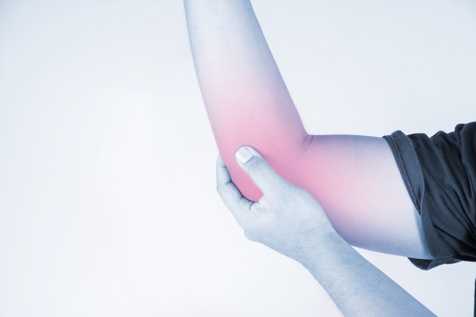
Coronoid fractures are uncommon but significant elbow injuries. The coronoid process is a key bony stabiliser of the elbow, and its fracture, particularly when associated with an elbow dislocation, can result in profound elbow instability requiring careful surgical reconstruction.
What is Coronoid Fracture?
The coronoid process is an anterior bony projection of the ulna that provides critical stability against posterior elbow dislocation. It also serves as the attachment point for the medial collateral ligament and the anterior capsule. Coronoid fractures most commonly occur as part of complex elbow injuries, including the terrible triad and posteromedial rotatory instability patterns.
Types of Coronoid Fracture
Type I
Regan-Morrey Type I (Tip Fracture): Only the tip of the coronoid process is fractured. These are most commonly seen in association with elbow dislocations. Often managed non-surgically when the elbow is otherwise stable after reduction.
Type II
Regan-Morrey Type II (Less than 50 percent of height): A fracture of the coronoid involving less than 50 percent of the coronoid height. Associated with greater instability and more commonly requires surgical fixation.
Type III
Regan-Morrey Type III (Greater than 50 percent of height): A large fracture involving more than 50 percent of the coronoid process. These fractures significantly destabilise the elbow and almost always require surgical repair.
Type IV
Anteromedial Facet Fracture (O'Driscoll Subtype): A specific fracture pattern involving the anteromedial corner of the coronoid, associated with posteromedial rotatory instability. Even small anteromedial facet fractures can produce significant varus-posteromedial instability and generally require surgical fixation.
Causes
Elbow dislocation is the most common associated event, with the coronoid fracturing as the ulna levers against it during the dislocation mechanism. Varus posteromedial rotatory stress, typically from a fall on the outstretched arm with a varus force applied to the elbow, specifically produces anteromedial facet fractures. High-energy trauma in road traffic accidents and sporting collisions can produce isolated coronoid fractures, though these are uncommon without an associated dislocation.
- Medial elbow pain and swelling following an elbow dislocation or significant elbow trauma
- Tenderness on palpation over the medial and anterior elbow
- Limited elbow flexion and extension, often with a sensation of instability particularly with attempts to straighten the elbow
- In posteromedial rotatory instability patterns: a sense of the elbow giving way or pivoting inward, particularly during pushing activities
- Associated ulnar nerve symptoms including tingling in the ring and little fingers

Diagnosis
Plain X-rays are the first investigation and may demonstrate the coronoid fracture, though small tip fractures and anteromedial facet fractures are often invisible on standard radiographs. CT scan with three-dimensional reconstruction is essential for all suspected coronoid fractures, as it accurately defines the fracture pattern, the degree of displacement, and the presence of associated radial head and ligamentous injuries.
MRI is valuable in assessing associated ligamentous injuries, particularly the medial and lateral collateral ligament complexes. Fluoroscopic examination under anaesthesia is used intraoperatively to confirm elbow stability.
Treatment
Surgical Fixation or Reconstruction
Significant coronoid fractures associated with elbow instability are treated surgically. Dr. Kushalappa repairs the coronoid fragment using screws, suture anchors, or bone grafting as appropriate to the fracture pattern, combined with repair of the associated ligamentous injuries and any radial head fracture in the same session.
Why Choose Dr. Kushalappa Subbiah in Bangalore?
Dr. Kushalappa Subbiah completed a Fellowship in Shoulder Surgery at the Sydney Shoulder Research Institute, where he trained in advanced arthroscopic shoulder procedures including labral repair and reconstruction. He holds the International Olympic Committee (IOC) Diploma in Sports Medicine, and has direct clinical experience managing shoulder injuries in Indian athletes across cricket, swimming, tennis, and contact sports. He is appointed as a Consultant Shoulder Surgeon at NH Hospital, Bangalore.
Frequently Asked Questions
Small tip fractures (Regan-Morrey Type I) without associated instability can often be managed non-surgically. Larger fractures, particularly those associated with elbow dislocation and multi-ligament injury, require surgical stabilisation.