6935 Todds Lane San Antonio, TX 98336
Our Working Time: 09:00 am To 06:00 pm

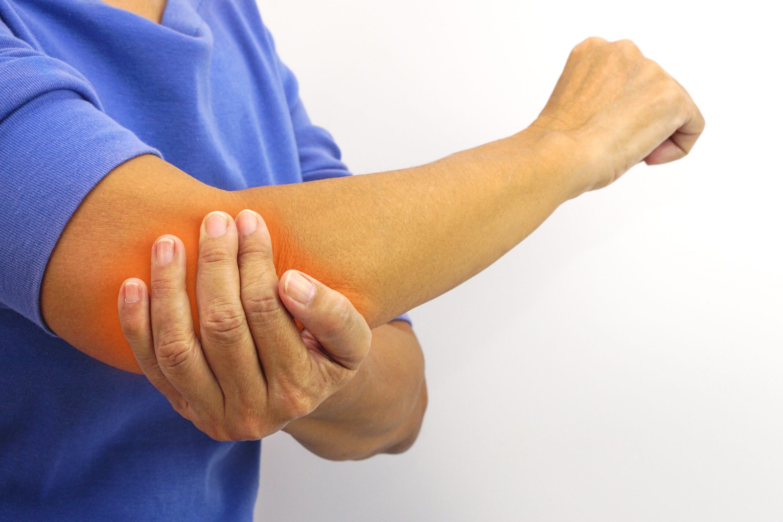
Progressive elbow pain, stiffness that makes it difficult to fully straighten or bend the arm, and a grinding or locking sensation in the joint are the hallmarks of elbow arthritis. While less common than knee or hip arthritis, it can be profoundly disabling when it affects the dominant arm of an active individual.
What is Elbow Arthritis?
Elbow arthritis refers to the deterioration of cartilage within the elbow joint, leading to progressive pain, loss of motion, and bony changes. The primary joint affected is the ulnohumeral articulation, and the radiohumeral and proximal radioulnar joints can also be involved. Common forms include osteoarthritis from progressive cartilage wear, rheumatoid arthritis as an autoimmune condition, and post-traumatic arthritis following previous elbow injuries or fractures.
Types of Elbow Arthritis
Type I
Primary Osteoarthritis: The most common form, caused by progressive cartilage wear and degeneration over time. More prevalent in males and in those who perform heavy manual work or repetitive loading of the elbow over many years. Characterised by osteophyte formation, loose bodies, and progressive loss of terminal flexion and extension.
Type II
Rheumatoid Arthritis: An autoimmune inflammatory condition that attacks the joint lining. Produces synovial hypertrophy, cartilage destruction, and eventual deformity. Often affects multiple joints and can affect patients of any age.
Type III
Post-Traumatic Arthritis: Develops as a consequence of previous elbow injury, most commonly after fractures, dislocations, or fracture-dislocations that damaged the articular cartilage or produced malunion. Often develops years after the original injury.
Type IV
Crystalline Arthropathy: Gout and pseudogout can affect the elbow joint, producing acute inflammatory episodes superimposed on chronic joint degeneration. Crystal deposition in the joint and surrounding soft tissues, including the olecranon bursa, is a characteristic finding.
Causes
Cumulative cartilage wear from years of heavy manual work, repetitive loading, or contact sport participation is the predominant cause of primary elbow osteoarthritis. Previous elbow fractures and dislocations that produced articular damage or post-traumatic instability are major risk factors for secondary arthritis. Autoimmune conditions, particularly rheumatoid arthritis, produce inflammatory destruction of the joint cartilage and synovium. Crystal deposition disorders such as gout contribute to progressive cartilage damage. Age is a consistent risk factor.
- Aching elbow pain worsening with activity and at rest in advanced stages
- Progressive loss of elbow flexion and extension, particularly difficulty fully straightening the arm
- A locking or catching sensation caused by loose bodies of bone or cartilage within the joint
- Swelling and tenderness around the elbow joint
- Pins and needles in the ring and little fingers in some patients, due to ulnar nerve irritation adjacent to the arthritic joint

Diagnosis
Plain X-rays demonstrate joint space narrowing, osteophyte formation, and loose bodies. CT scan provides better assessment of the bony anatomy and loose body location. MRI assesses remaining cartilage and soft tissue involvement.
Treatment
Elbow Arthroscopy
For early to moderate elbow arthritis with loose bodies and limited contracture, Dr. Kushalappa performs elbow arthroscopy. Loose bodies are removed, osteophytes are debrided, and any associated soft tissue contracture is released. This significantly reduces pain, improves elbow range of motion, and delays the need for joint replacement in appropriate patients.
Elbow Replacement (Total Elbow Arthroplasty)
For advanced elbow arthritis with severe pain and functional loss that has not responded to conservative management or arthroscopic treatment, Dr. Kushalappa performs total elbow arthroplasty. The damaged surfaces of the elbow joint are replaced with linked metal and polyethylene components, reliably relieving pain and restoring a functional range of elbow motion.
Why Choose Dr. Kushalappa Subbiah in Bangalore?
Dr. Kushalappa Subbiah completed a Fellowship in Shoulder Surgery at the Sydney Shoulder Research Institute, where he trained in advanced arthroscopic shoulder procedures including labral repair and reconstruction. He holds the International Olympic Committee (IOC) Diploma in Sports Medicine, and has direct clinical experience managing shoulder injuries in Indian athletes across cricket, swimming, tennis, and contact sports. He is appointed as a Consultant Shoulder Surgeon at NH Hospital, Bangalore.
Frequently Asked Questions
Elbow replacement is less commonly performed than hip or knee replacement, partly because the elbow tolerates load restrictions better than lower limb joints. It is most commonly indicated for inflammatory arthritis such as rheumatoid arthritis, and for advanced post-traumatic arthritis in the elderly.
In many patients with early to moderate elbow arthritis, arthroscopic debridement and loose body removal provides significant symptom relief lasting several years, potentially delaying or avoiding the need for joint replacement.