6935 Todds Lane San Antonio, TX 98336
Our Working Time: 09:00 am To 06:00 pm

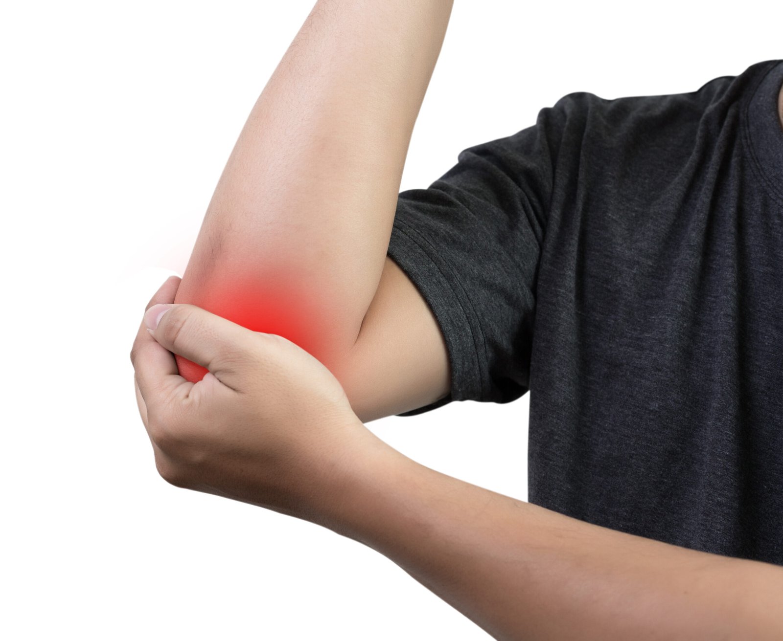
A fall on an outstretched arm producing immediate lateral elbow pain, difficulty rotating the forearm, and tenderness directly over the outer side of the elbow is the classic presentation of a radial head fracture. It is one of the most common elbow fractures in adults.
What is Radial Head Fracture?
Heterotopic ossification (HO) is the formation of mature bone in an abnormal location within soft tissues, typically around a joint. In the elbow, it most commonly develops following fractures, dislocations, burns, or neurological injuries such as head injury or spinal cord injury. The ectopic bone progressively restricts movement as it matures.
Types of Radial Head Fracture
Type I
Mason Type I: An undisplaced or minimally displaced fracture of the radial head. The articular surface is intact and forearm rotation is not mechanically blocked. Managed conservatively with early mobilisation and physiotherapy.
Type II
Mason Type II: A marginal sector fracture of the radial head with displacement greater than 2 millimetres or angulation, producing a mechanical block to forearm rotation. Surgical fixation with headless compression screws is appropriate when the fragment is large enough and of good bone quality.
Type III
Mason Type III: A comminuted fracture involving the entire radial head, not amenable to fixation. Radial head replacement with a metallic prosthesis is the treatment of choice.
Type IV
Mason Type IV: Any radial head fracture associated with an elbow dislocation. The combination of the fracture with ligamentous disruption makes this a highly unstable injury pattern requiring surgical management of both the fracture and the ligaments.
Type V
Essex-Lopresti Injury: A specific associated injury pattern in which the radial head fracture is accompanied by disruption of the interosseous membrane of the forearm and injury to the distal radioulnar joint. Radial head replacement is essential in this pattern to prevent proximal migration of the radius.
Causes
A fall on an outstretched arm is the most common mechanism, occurring in all age groups. The axial compressive force transmitted through the wrist and forearm is concentrated at the radial head as it impacts the capitellum. Direct impact to the lateral elbow from a contact sport collision or road traffic accident can also fracture the radial head. An elbow dislocation event that reduces spontaneously can leave behind a radial head fracture that is only identified on subsequent imaging.
- Immediate pain and tenderness localised to the lateral elbow, directly over the radial head
- Swelling around the lateral and posterior elbow developing within hours of the injury
- A painful restriction of forearm rotation, particularly pronation and supination
- Limited elbow flexion and extension due to pain and haemarthrosis within the joint
- In Essex-Lopresti injuries: wrist pain and distal radioulnar joint tenderness alongside the elbow symptoms

Diagnosis
Plain X-rays of the elbow in anteroposterior and lateral projections are the initial investigation. The radiocapitellar view provides improved visualisation of the radial head. A positive fat pad sign, where the posterior fat pad is elevated from the olecranon fossa, is a reliable indicator of haemarthrosis and should raise suspicion of an occult radial head fracture even when the fracture line is not directly visible.
CT scan is used for Mason Type II fractures where surgical planning is being considered, and for all Type III and Type IV injuries. MRI is reserved for cases where significant ligamentous injury is suspected alongside the fracture.
Treatment
Non-Surgical Management
Once the heterotopic bone has matured, typically 12 to 18 months following the original injury, Dr. Kushalappa performs surgical excision of the ectopic bone combined with elbow contracture release. Timing of surgery is important, as operating on immature HO is associated with a higher recurrence rate. Post-operative prophylaxis with anti-inflammatory medication or low-dose radiation is used to reduce the risk of recurrence.
Surgical Excision with Release
Once the heterotopic bone has matured, typically 12 to 18 months following the original injury, Dr. Kushalappa performs surgical excision of the ectopic bone combined with elbow contracture release. Timing of surgery is important, as operating on immature HO is associated with a higher recurrence rate. Post-operative prophylaxis with anti-inflammatory medication or low-dose radiation is used to reduce the risk of recurrence.
Why Choose Dr. Kushalappa Subbiah in Bangalore?
Dr. Kushalappa Subbiah completed a Fellowship in Shoulder Surgery at the Sydney Shoulder Research Institute, where he trained in advanced arthroscopic shoulder procedures including labral repair and reconstruction. He holds the International Olympic Committee (IOC) Diploma in Sports Medicine, and has direct clinical experience managing shoulder injuries in Indian athletes across cricket, swimming, tennis, and contact sports. He is appointed as a Consultant Shoulder Surgeon at NH Hospital, Bangalore.
Frequently Asked Questions
Yes, recurrence is a recognised risk, particularly if surgery is performed before the HO has fully matured. Post-operative prophylaxis, either with NSAIDs or low-dose radiation therapy, reduces but does not eliminate the risk of recurrence.