6935 Todds Lane San Antonio, TX 98336
Our Working Time: 09:00 am To 06:00 pm

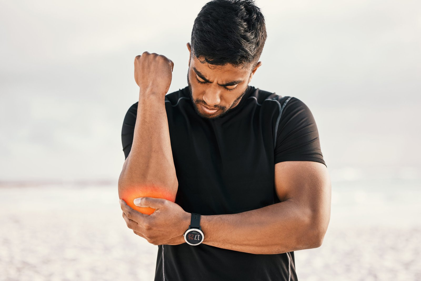
A capitellar fracture is a relatively uncommon but technically demanding elbow injury involving the rounded lateral portion of the distal humerus. It typically requires surgical fixation to restore the articular surface and prevent long-term elbow stiffness and arthritis.
What is Capitellar Fracture?
The capitellum is the rounded portion of the lateral distal humerus that articulates with the radial head. Capitellar fractures typically occur from a shearing force transmitted through the radial head during a fall. They are frequently associated with radial head fractures. Because the capitellum is almost entirely cartilage-covered with limited soft tissue attachment, surgical fixation requires precise technique.
Types of Capitellar Fracture
Type I
Hahn-Steinthal Fracture: A large osteochondral fracture of the capitellum, often involving a substantial portion of the articular surface. The fragment is typically displaced anteriorly and superiorly and usually contains sufficient bone for fixation with headless compression screws.
Type II
Kocher-Lorenz Fracture: A smaller fracture involving primarily the articular cartilage with minimal underlying subchondral bone. The thin, fragile nature of these fragments makes internal fixation technically challenging, and fragment excision may be required if fixation is not possible.
Type III
Comminuted Fracture: A severely comminuted capitellar fracture with multiple small fragments that cannot be anatomically reconstructed or fixed. Fragment excision is the typical treatment.
Type IV
Extended Lateral Column Fracture: A fracture that extends medially to involve the lateral trochlea, in addition to the capitellum. These fractures involve a larger portion of the distal humerus articular surface and are more complex to fix.
Causes
A fall on an outstretched arm transmitting an axial compressive load through the radial head into the capitellum is the most common mechanism. The shearing force created as the radial head impacts the capitellum during the fall cleaves the capitellar fragment from the rest of the distal humerus. Capitellar fractures are more common in women than men, which may be related to differences in elbow carrying angle that concentrate stress at the lateral elbow.
- Immediate lateral elbow pain and swelling following the injury, often mistaken initially for a simple elbow bruise or radial head fracture
- Painful restriction of elbow flexion, particularly beyond 90 degrees, as the displaced capitellar fragment mechanically blocks further motion
- Restriction of forearm rotation due to the close relationship between the capitellar fragment and the radial head
- Tenderness localised to the lateral elbow, slightly proximal to the radial head, over the capitellum
- A palpable click or crepitus during elbow movement if a fragment is displaced into the joint

Diagnosis
Plain X-rays, particularly the lateral elbow view, demonstrate a double arc sign where the outline of the displaced capitellar fragment can be seen overlying the distal humerus. However, capitellar fractures are frequently missed on initial plain radiographs, particularly smaller Type II fragments.
CT scan is essential for all suspected capitellar fractures, providing precise characterisation of the fracture type, fragment size, degree of comminution, and extension into the trochlea. This information directly determines whether fixation, excision, or a combined approach is planned. MRI is used when osteochondral injury without visible fracture is suspected.
Treatment
Open Reduction and Internal Fixation
Dr. Kushalappa performs anatomic reduction and fixation of capitellar fractures using headless compression screws inserted from anterior to posterior through the cartilage surface. When the capitellum is too comminuted to reconstruct, fragment excision may be considered, with careful assessment of the resulting elbow stability.
Why Choose Dr. Kushalappa Subbiah in Bangalore?
Dr. Kushalappa Subbiah completed a Fellowship in Shoulder Surgery at the Sydney Shoulder Research Institute, where he trained in advanced arthroscopic shoulder procedures including labral repair and reconstruction. He holds the International Olympic Committee (IOC) Diploma in Sports Medicine, and has direct clinical experience managing shoulder injuries in Indian athletes across cricket, swimming, tennis, and contact sports. He is appointed as a Consultant Shoulder Surgeon at NH Hospital, Bangalore.
Frequently Asked Questions
Untreated or malunited capitellar fractures lead to progressive elbow arthritis, loss of rotation, and potential lateral elbow instability due to the close relationship between the capitellum and the radial head. Prompt surgical treatment gives the best long-term outcome.