6935 Todds Lane San Antonio, TX 98336
Our Working Time: 09:00 am To 06:00 pm

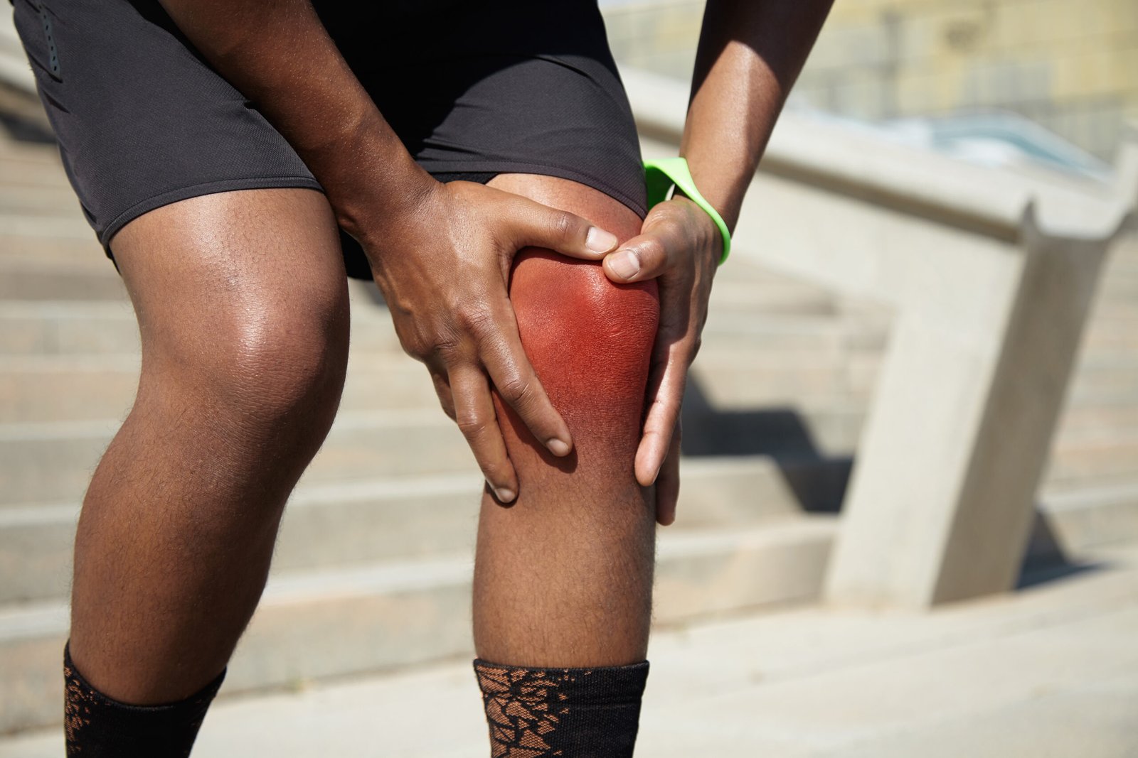
A kneecap that dislocates or slides sideways out of its groove, leaving the knee painful, swollen, and giving way during activity, is the experience of patellar instability. It is a condition that primarily affects younger patients, particularly females, and tends to recur without appropriate surgical management.
What is Patellar Instability?
The patella sits in the trochlear groove of the femur and is held in position by the quadriceps muscle mechanism, the patella tendon, and the medial patellofemoral ligament (MPFL) on the inner side. When the patella dislocates, it typically shifts laterally, tearing the MPFL. Without surgical repair or reconstruction of the MPFL, the risk of recurrent dislocation is high, particularly in patients with underlying anatomical risk factors such as a shallow trochlear groove or laterally positioned tibial tubercle.
Types of Patellar Instability
Type I
Acute First-Time Dislocation: The first episode of lateral patellar dislocation, typically in a young patient following a specific traumatic event such as a twisting movement, a sporting collision, or a direct blow to the medial knee. The MPFL is torn and the patient presents with acute pain, a haemarthrosis, and either a visible displaced patella or a recently self-reduced kneecap.
Type II
Recurrent Patellar Dislocation: Repeated episodes of lateral patellar dislocation following the initial event. Common in patients with underlying anatomical risk factors who have not undergone surgical stabilisation after their first dislocation. Each subsequent episode progressively damages the articular cartilage of the patella and trochlea, increasing the long-term risk of patellofemoral arthritis.
Type III
Chronic Patellar Subluxation: A pattern in which the patella does not fully dislocate but repeatedly shifts laterally during activity, producing pain, apprehension, and giving way without a complete dislocation event. Often associated with patella alta, trochlear dysplasia, or an increased tibial tubercle to trochlear groove (TT-TG) distance. Patients frequently describe a sense that the kneecap is about to go out rather than a full dislocation.
Type IV
Patellofemoral Pain Syndrome with Lateral Maltracking: A related but distinct condition in which the patella tracks laterally and unevenly through the trochlear groove during knee flexion and extension without frank dislocation or subluxation. Produces anterior knee pain with activity and prolonged sitting, and is often the precursor to more significant instability if the underlying anatomical factors are not addressed.
Causes
Anatomical risk factors are the most important predisposing element and include trochlear dysplasia, where the trochlear groove is shallow, flat, or convex rather than concave; patella alta, where the kneecap sits abnormally high above the groove and enters it late during knee flexion; and a lateralised tibial tubercle producing an increased TT-TG distance. Patients with these structural features require significantly less traumatic force to dislocate the patella and are at high risk of recurrence without surgical correction.
Generalised ligamentous laxity, which is more prevalent in females and in patients with connective tissue hypermobility conditions, reduces the passive restraint to lateral patellar migration and is a recognised risk factor for first-time and recurrent dislocation.
A specific traumatic event, such as a twisting injury, a direct blow to the medial aspect of the kneecap, or a sudden change of direction that places valgus stress on the knee, can cause dislocation even in patients without significant underlying anatomical risk factors, particularly in contact and court sports.
Weakness or inhibition of the vastus medialis oblique (VMO) portion of the quadriceps muscle reduces the dynamic medial restraint on the patella and allows progressive lateral shift during activity, contributing to both subluxation and frank dislocation.
- The kneecap visibly or palpably shifts out of its groove to the outer side of the knee
- Immediate pain and swelling following dislocation
- A feeling of giving way or apprehension when the knee is slightly flexed and the quadriceps are engaged
- Tenderness along the medial aspect of the patella where the MPFL has torn

Diagnosis
Clinical examination assesses patellar tracking and the presence of apprehension. The patellar apprehension test, in which gentle lateral pressure applied to the patella with the knee in slight flexion produces patient guarding and distress, is highly specific for patellar instability. Medial patellar tenderness at the MPFL attachment site on the medial femoral condyle and the medial patellar border is assessed. The J-sign, where the patella tracks smoothly through flexion but jumps sharply laterally as the knee approaches full extension, indicates significant lateral maltracking. Patellar tilt and medial and lateral glide are also assessed to quantify the degree of lateral retinacular tightness and medial restraint insufficiency.
Plain X-rays in the axial or skyline view demonstrate the relationship of the patella to the trochlear groove and may reveal a shallow or absent groove, a laterally tilted patella, or a small osteochondral fragment from the medial patellar facet produced by the dislocation event. CT scan with the knee in extension provides precise measurement of the TT-TG distance and detailed assessment of trochlear morphology, both of which are essential for surgical planning when tibial tubercle osteotomy is being considered alongside MPFL reconstruction. MRI assesses MPFL integrity and the degree of tearing, articular cartilage damage on the patella and trochlear groove surfaces, bone bruising patterns on the lateral femoral condyle and medial patellar facet characteristic of dislocation, and any associated soft tissue or meniscal injuries.
Treatment
MPFL Reconstruction or Repair
For recurrent patellar dislocation or first-time dislocation in patients with significant anatomical risk factors, surgical stabilisation through MPFL repair or reconstruction is recommended. Using arthroscopic and open techniques, the medial patellofemoral ligament is reconstructed using a tendon graft, restoring the medial restraint that prevents lateral patellar dislocation. In patients with a significantly lateralised tibial tubercle contributing to instability, a tibial tubercle osteotomy to reposition the attachment of the patella tendon is combined with the MPFL reconstruction.
Why Choose Dr. Kushalappa Subbiah in Bangalore?
Dr. Kushalappa Subbiah completed a Fellowship in Shoulder Surgery at the Sydney Shoulder Research Institute, where he trained in advanced arthroscopic shoulder procedures including labral repair and reconstruction. He holds the International Olympic Committee (IOC) Diploma in Sports Medicine, and has direct clinical experience managing shoulder injuries in Indian athletes across cricket, swimming, tennis, and contact sports. He is appointed as a Consultant Shoulder Surgeon at NH Hospital, Bangalore.
Frequently Asked Questions
Not always. A first-time dislocation in a patient without significant anatomical risk factors can be treated with physiotherapy and quadriceps rehabilitation with a reasonable success rate. However, younger patients, females, and those with anatomical risk factors (shallow trochlea, high patella, lateralised tibial tubercle) have a high recurrence rate and are often better served with early surgical stabilisation.
Yes. MPFL reconstruction has a very high success rate at preventing further dislocations. Combined with a thorough physiotherapy programme to restore quadriceps strength and neuromuscular control, the vast majority of patients do not experience further instability after surgical stabilisation.