6935 Todds Lane San Antonio, TX 98336
Our Working Time: 09:00 am To 06:00 pm

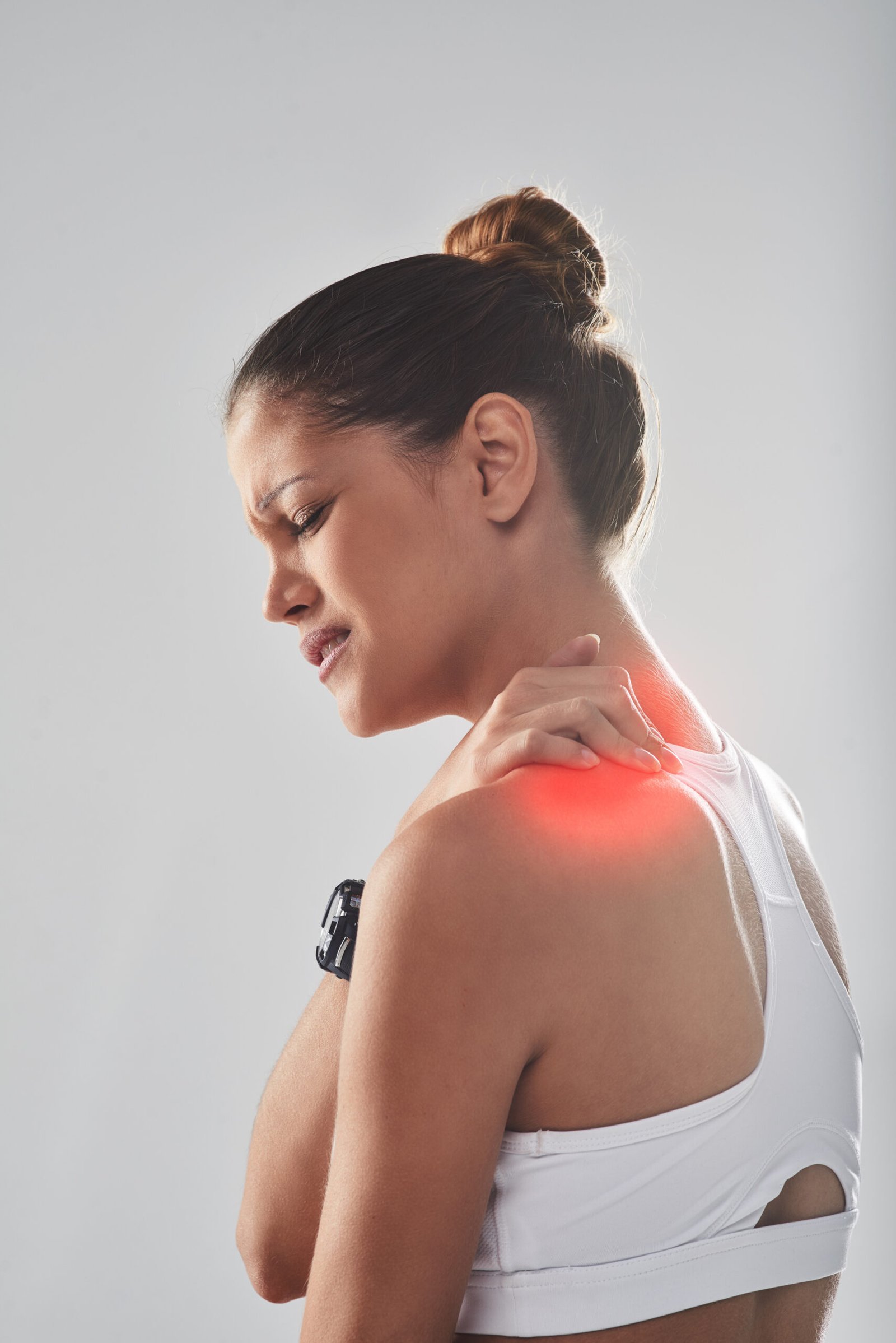
A sharp pain at the very top of the shoulder following a fall or direct blow, sometimes accompanied by a visible bump where the collarbone meets the shoulder blade, is one of the most characteristic presentations in sports and orthopaedic practice. Acromioclavicular joint injuries are among the most common shoulder injuries seen in contact and overhead sports, and the management decision between conservative and surgical treatment depends heavily on the degree of disruption.
Dr. Kushalappa Subbiah provides specialist assessment and surgical reconstruction of AC joint injuries and instability at his Bangalore clinics, with dedicated experience in managing these injuries in active patients and athletes.
What is AC Joint?
The acromioclavicular (AC) joint is the articulation between the outer end of the clavicle (collarbone) and the acromion process of the scapula (shoulder blade). It sits at the very top of the shoulder and is stabilised by two sets of ligaments: the acromioclavicular ligaments, which provide horizontal stability across the joint, and the coracoclavicular ligaments (the trapezoid and conoid), which provide vertical stability by suspending the scapula from the clavicle.
When the AC joint is injured, these ligaments are stretched or torn, allowing the clavicle to separate from the acromion. This separation can be mild, with the joint remaining essentially stable, or complete, with the clavicle riding visibly above its normal position.
Types of SC Joint Conditions
Type I
Type I: Sprain of the acromioclavicular ligaments only. The joint remains stable and in normal alignment. No visible deformity. Treated conservatively.
Type II
Type II: Complete tear of the acromioclavicular ligaments with a partial tear of the coracoclavicular ligaments. Mild widening of the joint on X-ray. A slight step deformity may be present. Generally managed conservatively.
Type III
Type III: Complete tear of both the acromioclavicular and coracoclavicular ligaments. The clavicle displaces upward, creating a clearly visible step deformity at the shoulder tip. The joint is unstable. Management is debated between conservative and surgical.
Type VI
Type VI: Rare inferior dislocation of the clavicle below the coracoid. Associated with significant neurovascular injury and requires surgical management.
Type V
Type V: Severe superior displacement of the clavicle due to stripping of the deltoid and trapezius muscle attachments. The step deformity is dramatic. Requires surgical reconstruction.
Causes
The most common mechanism is a direct fall onto the tip of the shoulder, typically from a bicycle, during a rugby tackle, or from a height. The force drives the acromion downward while the clavicle is held in place by its muscular attachments to the neck, creating a separation force across the AC joint. A fall onto an outstretched arm is a secondary mechanism. AC joint injuries are common in cricket, cycling, rugby, football, kabaddi, and hockey.
- Pain directly at the top of the shoulder, sharply localised over the AC joint, worsening with overhead movement, cross-body adduction, and lying on the affected side
- A visible bump or step deformity at the shoulder tip in Type III and above, representing the displaced clavicle
- Tenderness on direct palpation of the AC joint
- Restricted shoulder movement, particularly painful when raising the arm across the body or above shoulder height
- Weakness with pushing or overhead activity in higher-grade injuries due to disruption of the muscle attachments
- In chronic cases: a sense of shoulder instability with heavy loading or overhead activities, and persistent aching at the joint

Diagnosis
Clinical examination of the SC joint is essential and includes careful assessment of the position of the medial clavicle, local tenderness, and any signs of posterior compression of mediastinal structures. Standard shoulder X-rays are often insufficient to assess the SC joint adequately. CT scan is the investigation of choice for characterising the degree of displacement, identifying associated fractures, and distinguishing anterior from posterior dislocation. MRI is used in chronic presentations where ligament integrity and the intra-articular disc need to be assessed.
Treatment
Non-Surgical Management
Type I and II injuries are managed conservatively with a sling for comfort, ice, progressive physiotherapy once pain allows, and a graduated return to sport. Type III injuries are a subject of ongoing debate in the surgical literature. The majority of patients with Type III injuries, particularly those with low-to-moderate functional demands, can be managed successfully without surgery using a structured rehabilitation programme. Dr. Kushalappa will discuss the evidence and the individual patient's activity level and goals when advising on Type III management.
Arthroscopic AC Joint Stabilisation and Reconstruction
Type IV, V, and VI injuries, and selected Type III injuries in high-demand athletes or patients who have failed conservative management, are treated surgically. Dr. Kushalappa performs arthroscopic-assisted AC joint stabilisation and coracoclavicular ligament reconstruction using tendon graft. The procedure restores the clavicle to its normal anatomical position and reconstructs the vertical stabilisers of the joint, allowing reliable return to sport and heavy overhead activity. Recovery involves a sling for 4 to 6 weeks, physiotherapy progressing from 2 weeks, and return to contact sport at 4 to 6 months.
Why Choose Dr. Kushalappa Subbiah in Bangalore?
Dr. Kushalappa Subbiah completed a Fellowship in Shoulder Surgery at the Sydney Shoulder Research Institute, where he trained in advanced arthroscopic shoulder procedures including labral repair and reconstruction. He holds the International Olympic Committee (IOC) Diploma in Sports Medicine, and has direct clinical experience managing shoulder injuries in Indian athletes across cricket, swimming, tennis, and contact sports. He is appointed as a Consultant Shoulder Surgeon at NH Hospital, Bangalore.
Frequently Asked Questions
No. Type I and II injuries heal well without surgery. Most Type III injuries also do well with conservative management. Surgery is recommended for Types IV, V, and VI, and for Type III injuries in patients with high functional demands where conservative treatment has not restored adequate shoulder function.
In higher-grade AC joint separations, the visible step deformity from the elevated clavicle often persists even after the pain has fully resolved. In patients managed conservatively, the bump remains but is typically not functionally significant. In surgically reconstructed patients, the clavicle is returned to its normal position and the deformity is corrected.
For conservatively managed Type I and II injuries, return to sport can occur within 2 to 4 weeks once pain allows. For Type III injuries managed conservatively, return to contact sport typically takes 6 to 12 weeks. Following surgical reconstruction, return to contact sport is expected at 4 to 6 months.
No. The AC joint is a separate structure at the very top of the shoulder where the clavicle meets the acromion. The rotator cuff is a group of four tendons inside the shoulder joint that attach the arm to the shoulder blade. Both structures can be injured simultaneously, and AC joint injuries can contribute to rotator cuff impingement.